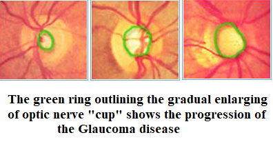
Glaucoma can be caused by increased pressure of the fluid in the eye. The inside of the eye contains fluid that is constantly flowing into and being drained out of the eye. If the drainage mechanism in an area called Trabecular meshwork gets blocked, fluid starts accumulating in the eye, exerting pressure inside the eye. This extra fluid that builds up in the eye presses against the optic nerve at the back of the eye, thus damaging parts of the optic nerve. This damage appears as gradual visual changes and then loss of vision, if it remains untreated.
Types of Glaucoma
The most common type of glaucoma, known as chronic open angle (COAG) or primary open angle, occurs when the canals draining the eye of aqueous humor become clogged. This blockage can gradually increase pressure within the eye to damaging levels. No pain occurs, so individuals are usually unaware that these changes are occurring. There are no early signs or symptoms but over the years vision will be lost starting in the periphery and moving towards the central vision.
When eye pressure builds up rapidly, it is called acute angle-closure glaucoma. This type of glaucoma commonly occurs in individuals who have narrow anterior chamber angles. In these cases, aqueous fluid behind the iris cannot pass through the pupil thus pushing the iris forward, preventing aqueous drainage through the angle. It is as though a sheet of paper floating near a drain suddenly drops over the opening and blocks the flow out of the sink. In cases of acute angle closure glaucoma, one may experience blurred vision, halos around lights, deep pain behind the eye, nausea, and vomiting.
Swaraashi advises its patients to have periodic eye examinations for early detection of glaucoma.
Risk Factors for Glaucoma
High pressure alone does not mean that you have glaucoma. In Nethradhama we put together many kinds of information to determine your risk for developing this disease. The most important risk factors include:
In Unique eye care, the glaucoma specialist will weigh all of these factors before deciding whether you need treatment for glaucoma, or whether you should be monitored closely as a glaucoma suspect. Being glaucoma suspect means your risk of developing glaucoma is higher than normal, and you need to have regular examination to detect the early signs of damage to the optic nerve.
Diagnostic Facility
Regular glaucoma check-up in UNIQUE eye care includes a detailed history and ocular examination.
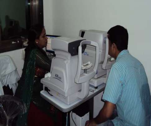
The technique of measurement of pressure within the eye (Intra ocular pressure – IOP/IOT) is called tonometry. This is very important since IOP is one of the indicators of Glaucoma. Tonometry is recommended in everyone over the age of 40, family history of glaucoma, Myopia, Diabetes and any history of previous ocular injuries. This technique can be performed with either applanation or non-contact tonometer.
Ophthalmoscopy
Ophthalmoscopy is used to examine the inside of the eye, especially the optic nerve. In a darkened room, the doctor will examine your eye by using an ophthalmoscope (an instrument with a small light on the end). This helps the doctor look at the shape and color of the optic nerve.If the pressure in the eye is not in the normal range, or if the optic nerve looks unusual, then one or two special glaucoma tests will be done. 3-Dimensional examination of optic disk too is done.
Visual Field Examination
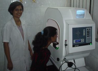
The perimetry test is also called a visual field test. During this test, you will be asked to look straight ahead and then indicate when a stationary light appears in the field of vision. This helps draw a "map" of your field of vision. This is a non-invasive computerised test which takes about 10-20 minutes to perform and gives information regarding optic nerve damage due to glaucoma.
Optical Coherence Tomography (OCT)
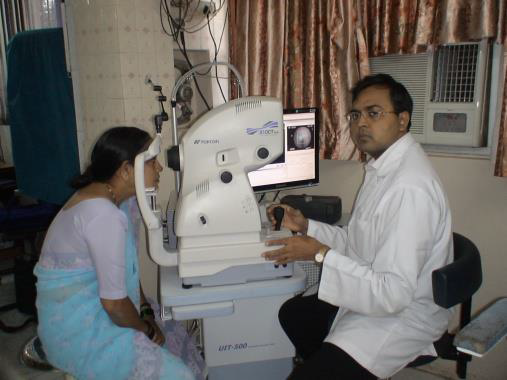
Optical coherence tomography is a new, noninvasive, noncontact, imaging technology capable of early detection of glaucoma through it's ability to evaluate the nerve cells damaged in glaucoma.
Glaucoma Services
Glaucoma is usually controlled with eye drops taken daily which prevent damage to the optic nerve by lowering eye pressure, either by slowing the production of aqueous fluid within the eye or by improving the flow out of the eye. Recently there have been a few new medications which show great promise for more effectively controlling eye pressure. In order for these medications to work, you must take them regularly and continuously as they are prescribed. Quite simply, the key to the success of medication therapy is patient compliance.
If topical and/or oral therapy is not controlling the intraocular pressure (IOP), or if the patient is not taking their medicine according to schedule, laser surgery treatment may be an effective alternative or adjunct. The laser is usually used in one of two ways. In open-angle glaucoma, the laser is used to enlarge the drain (Diode laser trabeculoplasty) to help control eye pressure. In angle-closure glaucoma, the laser creates a hole in the iris
( Yag Laser Iridotomy ) to improve the out flow of aqueous fluid to the drain.
Glaucoma is a highly misunderstood disease. Frequently, patients do not even realize that they are affected by the disease and rarely have an inkling about its severity. Here are the most common myths and truths about glaucoma:
Everyone is at risk for glaucoma from babies to elderly people. But, older people are at a higher risk for glaucoma. However babies (approximately 1 out of every 10,000 babies are born with glaucoma) and young adults can also get glaucoma.
Glaucoma is not curable, however, it is manageable. But first it must be diagnosed. Often glaucoma can be managed with medication and/or surgery. This means that further loss of vision may be halted. However, glaucoma is a chronic disease that must be treated for lifetime.
With open angle glaucoma, the most common form, there are virtually no symptoms. There is usually no pain involved with the rise in eye pressure. Loss of vision begins with peripheral or side vision. This type of vision loss can be easily compensated for (by turning the head to the side) and may not be noticed until significant vision is lost. The best way to protect your sight from glaucoma is to be tested so that if you have glaucoma, treatment can begin immediately.
Glaucoma can infact cause blindness if it is left untreated. And unfortunately, many patients do not realise that are afflicted with glaucoma until they have lost nearly all vision.
Cure And Care Glaucoma At Swaraashi.
There is no cure. However, Glaucoma can be controlled if detected early. Appropriate and timely treatment goes a long way in either slowing down or halting the progression of the disease.
As there are no warning signs or symptoms, don't wait for visual problems to develop but get a thorough checkup at the age of 35 yrs, another at 40 yrs and every year there after in Unique eye care.
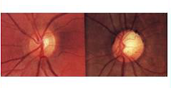
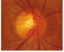
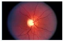
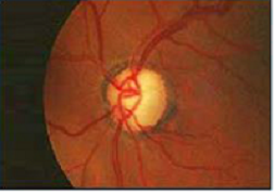
The photographs below demonstrate various degrees of optic nerve head cupping caused by increased intraocular pressure
